Micrograph Library
Browse the libraryAdvanced searchSystemsCompositionsTechniquesKeywordsPhase diagramsHelpPreferencesAbout the micrograph libraryTerms of useContribute micrographs!FeedbackLinksCredits Print this page
Full Record for Micrograph 581
[112 KB]
View micrograph
.. in new window
View micrograph and record
.. in new window
You can also view and download the micrographs on Flickr
- Micrograph no
- 581
- Brief description
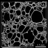
- X-ray tomography image of open cell polyurethane foam
- Keywords
- cell
 , composite material , foam , polymer , polymer composite foam, polyurethane (PU), thermoset, X-ray tomography
 - Categories
- Composite, Foam, Polymer, Polymer composite foam
- System
- Polyurethane (PU)
- Composition
- Not specified
- Standard codes
- Reaction
- Processing
- The principle of x-ray tomography is to re-construct the spatial distribution of the linear attenuation coefficient within the object from X-ray radiographs recorded at different angular settings.
- Applications
- This open celled polymer foam is a compliant space-filler, used for padding and in furniture. It may also be used as a precursor in processes such as the two-stage casting of a metal foam.
- Sample preparation
- Technique
- X-ray tomography
- Length bar
- 1.5 mm
- Further information
- If a gas is injected into a liquid it forms a cellular foam structure. When a thermoset prepolymer of low viscosity is foamed, the polymer can drain from the cell walls (driven by surface tension) before it sets at the cell edges, leaving an open-celled foam. The cell edges have three concave sides. The average co-ordination number for the nodes (where struts meet) is four, giving tetrahedral junctions. The deformation behaviour of the foam was observed by X-ray microtomography at the ESRF in Grenoble. X-ray tomography overcomes the limitations of MRI in terms of contrast, and those of confocal optical microscopy in terms of depth of field.
- Contributor
- Dr J A Elliott
- Organisation
- Department of Materials Science and Metallurgy, University of Cambridge
- Date
- 03/10/02
- Licence for re-use
 Attribution-NonCommercial-ShareAlike 4.0 International
- Related micrographs